

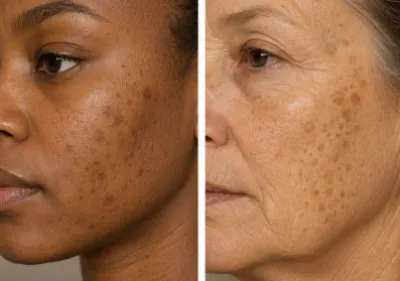
Dark patches, uneven skin tone, marks left behind by spots - these are among the most common skin concerns in the UK, and yet the term "hyperpigmentation" is frequently used as though it describes a single condition with a single solution. It does not. Hyperpigmentation covers a wide range of distinct presentations, each with its own cause, depth, and response to treatment. Using the wrong approach for the wrong type can slow progress, and in some cases, make things worse.
Part of the confusion is understandable. Melasma, post-inflammatory hyperpigmentation, sun spots, and freckles can all look broadly similar, particularly in photographs or on a phone screen. They share the same visible outcome - darker areas of skin - but they arise through different biological processes. A hormonal patch and a sun-damaged spot are not the same thing, even if they sit next to each other on the same cheek.
The depth at which pigment sits in the skin matters too. Brown marks that sit in the upper layers tend to respond more predictably to treatment. Grey or blue-grey marks that sit deeper are considerably more resistant, and some may be linked to medication rather than to sun exposure or inflammation. These distinctions are not widely explained in consumer-facing content, which is one reason people often cycle through products and procedures without a clear sense of why results vary.
This guide works through each type of hyperpigmentation in plain terms - what causes it, how to recognise it, and what the evidence suggests about managing it.
Starting with the basics: what hyperpigmentation actually is, and why the underlying mechanism matters for understanding everything that follows.
Our Preferred Partner - LaserMe by Neauvia
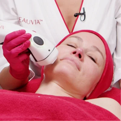
LaserMe is a non-ablative fractional diode laser used in medical aesthetics clinics across the UK to support skin regeneration and pigmentation management. This guide is for anyone trying to understand what it is, how it works, and whether it may be relevant to their hyperpigmentation concern.
- What LaserMe is, and how it differs from ablative laser treatments
- Which pigmentation concerns it is commonly used for, including PIH, melasma, and sun damage
- What to consider around suitability, recovery, costs, and choosing a qualified practitioner
What Is Hyperpigmentation?
Hyperpigmentation is a broad term used to describe areas of skin that have become darker than the surrounding tissue. It is not a single condition - it is a category that covers a wide range of causes and appearances, all connected by the same underlying process: the overproduction of melanin.
Why does hyperpigmentation happen?
Melanin is the pigment responsible for skin, hair, and eye colour. It is produced by cells called melanocytes, which sit in the deepest layer of the outer skin. In most circumstances, melanocytes produce melanin at a steady, regulated rate. When something disrupts that process - whether UV exposure, inflammation, a hormonal shift, or a medication - melanocytes can go into overdrive, depositing excess pigment in concentrated areas.
The result is visible darkening. This can range from a small, defined spot to a larger, more diffuse patch, depending on the cause and the individual's skin type.
Is hyperpigmentation dangerous?
In the vast majority of cases, hyperpigmentation is harmless. It causes no physical discomfort and poses no direct health risk. That said, some presentations - particularly those that appear suddenly, change in shape, or develop alongside other symptoms - can occasionally signal an underlying condition that warrants medical attention. Most dark patches, however, are cosmetic in nature and respond to appropriate treatment over time.
Understanding what type of hyperpigmentation you are dealing with is the most useful first step, both for setting realistic expectations and for choosing the right approach.
The Main Types Of Hyperpigmentation
Hyperpigmentation is not a single condition with a single cause. Several distinct types exist, each with different triggers, appearances, and responses to treatment. Knowing which type you have matters - not just for choosing the right product or procedure, but for understanding what realistic improvement looks like.
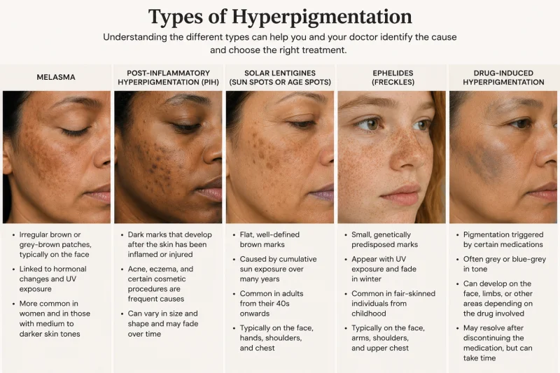
The most commonly seen types are:
Melasma - irregular brown or grey-brown patches, typically on the face, linked to hormonal changes and UV exposure. More common in women and in those with medium to darker skin tones.
Post-inflammatory hyperpigmentation (PIH) - dark marks that develop after the skin has been inflamed or injured. Acne, eczema, and certain cosmetic procedures are frequent causes.
Solar lentigines (sun spots or age spots) - flat, well-defined brown marks caused by cumulative sun exposure over many years. Common in adults from their 40s onwards.
Ephelides (freckles) - small, genetically predisposed marks that appear with UV exposure and fade in winter. Common in fair-skinned individuals from childhood.
Drug-induced hyperpigmentation - pigmentation triggered by certain medications, often grey or blue-grey in tone, which can develop on the face, limbs, or other areas depending on the drug involved.
There are rarer types too, including pigmentation linked to systemic conditions such as Addison's disease, and forms associated with chronic skin disorders. These fall outside the cosmetic category and are covered briefly in the section on when to seek professional advice.
Each type has a different underlying mechanism, which is why treatments that work well for one can have little effect - or occasionally worsen - another. A comparison table of key characteristics follows below.
| Type | Typical appearance | Main trigger | Tends to fade naturally? |
|---|---|---|---|
| Melasma | Brown or grey-brown patches | Hormones, UV | Slowly, with sun protection |
| PIH | Brown or blue-grey marks | Inflammation, injury | Often yes, over months |
| Solar lentigines | Small flat brown spots | Cumulative UV | No |
| Freckles | Small reddish-brown spots | UV, genetics | Yes, in winter |
| Drug-induced | Grey or blue-grey patches | Medication | Slowly, after stopping drug |
What Is Melasma?
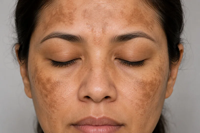
Melasma is one of the most commonly searched types of hyperpigmentation, and also one of the most frequently misunderstood. It presents as irregular patches of light to dark brown, or sometimes grey-brown, discolouration - typically appearing on the cheeks, forehead, upper lip, and jawline. Unlike a sun spot or a post-acne mark, melasma tends to develop across broader areas and usually appears on both sides of the face symmetrically.
What causes melasma?
Hormonal changes are the primary driver. Melasma is strongly associated with pregnancy, use of the combined contraceptive pill, and hormone replacement therapy - which is why it is sometimes called the "mask of pregnancy." That said, it can develop in anyone, including men, and UV exposure plays a significant role in triggering and worsening it even when hormones are the root cause. The two factors tend to work together: hormones sensitise the melanocytes, and sunlight activates them.
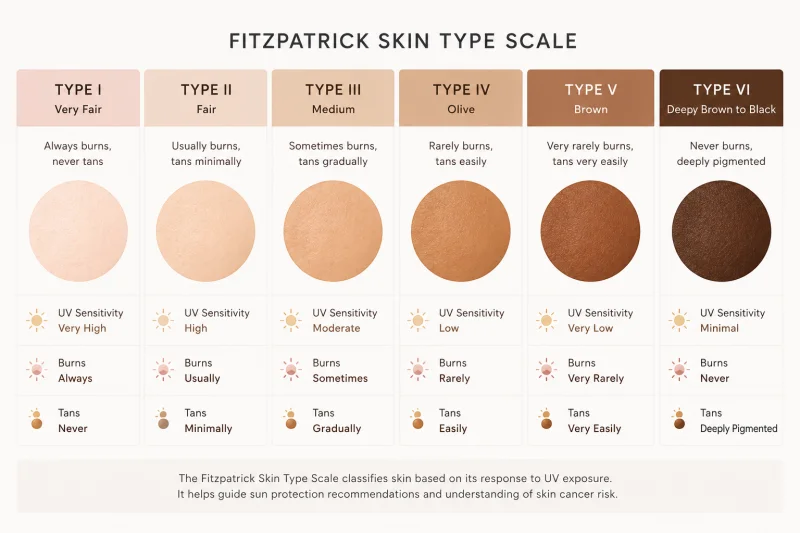
Genetics also has a part to play. Melasma is more common in those with a family history of the condition, and it occurs more frequently in people with Fitzpatrick skin types III to VI - broadly, medium to darker skin tones.
Why does melasma keep coming back?
This is one of the most common frustrations for people managing melasma. Even after successful treatment, it can return - sometimes within weeks of sun exposure. That is because melasma does not have a single, switchable cause. The melanocytes in affected areas remain sensitised, meaning ongoing sun protection is not optional; it is the foundation of any long-term management plan.
Epidermal and dermal melasma
Not all melasma sits at the same depth in the skin, and this affects how it responds to treatment. Epidermal melasma appears brown with relatively well-defined edges and tends to respond more readily to topical treatments. Dermal melasma sits deeper, appears grey-brown with less distinct margins, and is considerably more resistant. Mixed types, involving both layers, are also common. A dermatologist can assess this using a Wood lamp, though colour and edge definition offer a useful rough guide.
Melasma that appears predominantly brown is generally more treatable than patches with a grey or blue-grey tone.
What Is Post-inflammatory Hyperpigmentation, And Is It The Same As An Acne Mark?
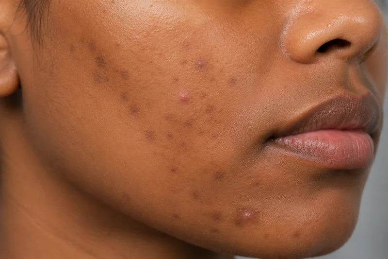
Post-inflammatory hyperpigmentation (PIH) is one of the most common types of hyperpigmentation, and one of the most frequently misnamed. Many people refer to the dark marks left after spots as acne scars, but in most cases, what they are seeing is PIH, a pigmentation change rather than a structural change to the skin. The distinction matters because the two respond to very different treatments.
What causes PIH?
PIH develops when the skin experiences inflammation or injury. During the inflammatory response, the body releases a cascade of chemical signals that stimulate melanocytes to produce excess melanin. Once the inflammation settles, that excess pigment remains - sometimes for months, sometimes longer.
Acne is a common trigger, but far from the only one. PIH can follow eczema flares, psoriasis, insect bites, burns, allergic reactions, and even certain cosmetic procedures, including chemical peels and laser treatments, if not appropriately matched to skin type. Any condition that causes significant inflammation can leave a pigmented mark.
Why does picking spots make marks worse?
This is worth addressing directly because it is a common behaviour with a clear consequence. Squeezing or picking a spot forces the inflammation deeper into the skin and prolongs it. The longer and more intense the inflammatory event, the more melanin is likely to be deposited - and the deeper it may sit. Mild acne that resolves on its own is considerably less likely to cause significant PIH than a lesion that has been repeatedly manipulated.
How is PIH different from acne scarring?
Acne scarring involves a physical change to the skin's structure - typically a depression (atrophic scar) or, less commonly, a raised area (hypertrophic scar). PIH involves no structural damage. The skin's surface remains smooth; only the colour has changed. In practice, both can be present simultaneously, which adds to the confusion. A trained practitioner can distinguish between them during a skin assessment.
Who is most affected by PIH?
PIH occurs across all skin types, but it is more common and more persistent in those with darker skin tones. Research indicates that in individuals with Fitzpatrick skin types IV to VI, acne-associated PIH can affect up to 65% of patients. Higher baseline melanin production and more reactive melanocytes mean the inflammatory response generates a stronger pigmentation response. That said, PIH can develop in lighter skin tones too - it simply tends to be less intense and fade more quickly.
PIH that appears brown is typically epidermal and has a reasonable chance of improving over time with appropriate care. Marks with a grey or blue-grey tone indicate deeper, dermal pigmentation - more persistent, and less responsive to topical treatments alone.
What Are Sun Spots And Age Spots?
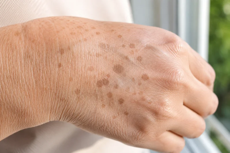
Sun spots - also known as age spots, liver spots, or by their clinical name solar lentigines - are flat, well-defined brown marks that develop on areas of skin exposed to the sun over many years. They are among the most common forms of hyperpigmentation in adults and, unlike some other types, do not fade on their own once formed.
What causes sunspots?
The cause is cumulative UV exposure. Over time, repeated sun exposure gradually increases melanin production in localised areas, eventually producing visible marks. This is a slow process - sun spots typically begin appearing from the 40s onwards, though they can develop earlier in those with significant sun exposure history or lighter skin tones. They are most common on the face, hands, shoulders, forearms, and chest: the areas that tend to receive the most unprotected sun over a lifetime.
Sun spots are distinct from a tan, which is a temporary, even response to UV exposure. A solar lentigo is a permanent localised change that does not reverse when sun exposure reduces.
How are sun spots different from freckles?
The two are often confused, but the distinction is straightforward. Freckles tend to be smaller, appear earlier in life, and fade noticeably in winter or with reduced sun exposure. Sun spots are larger, more defined, appear later in life, and do not fade seasonally. Both are benign, but solar lentigines are considerably more persistent and are generally the focus of treatment in adults seeking to address uneven skin tone.
When should sun spots be checked?
Most solar lentigines are harmless and require no medical treatment. However, any mark that changes in size, shape, or colour - or that develops an irregular border, multiple tones, or begins to raise above the skin surface - should be assessed by a qualified clinician. A flat brown spot and an early melanoma can sometimes look similar to an untrained eye. When in doubt, a professional assessment is always the right step.
What Are Freckles, And Do They Count As Hyperpigmentation?
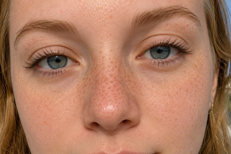
Freckles are a form of hyperpigmentation, though they sit at the more benign end of the spectrum and rarely require treatment. Technically known as ephelides, they are small, reddish to light-brown spots that develop on sun-exposed areas of skin - most commonly the face, arms, and shoulders. They are particularly common in fair-skinned individuals and tend to appear from childhood onwards.
What causes freckles?
Freckles are primarily genetic. People with fair skin and lighter hair are more likely to develop them, due to inherited differences in how their melanocytes respond to UV exposure. Sun exposure does not create new melanocytes in these areas - it simply activates the ones already present, causing them to produce more melanin temporarily. This is why freckles become more prominent in summer and fade in winter, when UV levels drop, and melanocyte activity reduces.
This seasonal fading is one of the key characteristics that separates freckles from other types of hyperpigmentation. A mark that reliably lightens in winter is very likely to be an ephelide rather than a solar lentigo or a PIH mark.
Are freckles the same as sun spots?
They are related but distinct. Both involve localised melanin production triggered by UV exposure, but the similarities largely end there. Freckles are smaller, appear earlier in life, have a genetic basis, and fade without sun exposure. Sun spots are larger, more defined, and develop later in life as a result of cumulative UV damage, and do not fade seasonally. In practice, adults sometimes find that marks they assumed were freckles have gradually become more fixed and defined over time - a sign that solar lentigines may be developing alongside or replacing the original ephelides.
Do freckles need treatment?
Freckles are benign and do not require medical treatment. Some individuals choose to address them for cosmetic reasons, in which case options such as topical brightening ingredients, chemical peels, and certain laser treatments may be discussed with a practitioner. That said, freckles often return with sun exposure, so realistic expectations and consistent sun protection are important parts of any conversation about treatment.
Can Medication Cause Hyperpigmentation?
Drug-induced hyperpigmentation is more common than most people realise, and it is almost entirely absent from consumer-facing skin content. For anyone who has developed unexplained dark or grey patches while taking long-term medication, this connection is worth understanding - because without knowing the cause, finding the right approach to managing it becomes considerably harder.

Which medications can cause hyperpigmentation?
Several widely used drugs are associated with pigmentation changes. These include:
Minocycline - an antibiotic commonly prescribed for acne. Long-term use can cause grey-blue discolouration, particularly on the face, legs, and areas of scarring.
The combined contraceptive pill - hormonal contraceptives can trigger or worsen melasma-type pigmentation in susceptible individuals, in a similar way to pregnancy-related hormonal changes.
Antimalarial drugs - including hydroxychloroquine and chloroquine, used for conditions such as lupus and rheumatoid arthritis as well as malaria prevention. These can cause blue-grey pigmentation on the face and lower legs.
Amiodarone - a heart medication associated with a distinctive grey or slate-blue discolouration on sun-exposed skin.
Tricyclic antidepressants - particularly imipramine, which can produce grey-brown pigmentation with prolonged use.
Certain chemotherapy agents - including busulfan and other cytotoxic drugs, which are associated with diffuse or localised darkening.
This is not an exhaustive list. If unexplained pigmentation develops during or after a course of medication, a GP or dermatologist is the right person to assess whether the drug could be a contributing factor.
What does drug-induced pigmentation look like?
Unlike the warm brown tones typical of sun damage or PIH, drug-induced pigmentation often has a grey, slate, or blue-grey quality. This reflects that many drug-related pigmentation changes occur deeper in the dermis rather than in the skin's surface layers. The grey tone is a practical indicator that the pigment sits deeper, which also explains why it tends to be more persistent and less responsive to standard topical treatments.
Will it improve if the medication is stopped?
In many cases, yes - but improvement is often slow. For some medications, it can take many months or even years for pigmentation to fade after the drug is discontinued. In some instances, particularly with minocycline or amiodarone, the change may be long-lasting. This is an important consideration and one that is best discussed with the prescribing clinician before making any decisions about stopping or changing medication.
Does The Depth Of Pigmentation Affect How Treatable It Is?
When it comes to hyperpigmentation, depth matters as much as type. Two people can have marks that look broadly similar on the surface but respond very differently to the same treatment - and the reason often comes down to where in the skin the pigment actually sits. Understanding this distinction helps set realistic expectations before starting any treatment course.
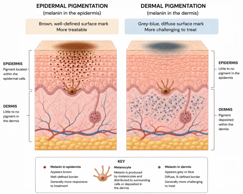
Epidermal pigmentation
Epidermal pigmentation sits in the upper layers of the skin. It typically appears brown, with relatively well-defined edges, and tends to respond more readily to treatment. Topical ingredients that inhibit melanin production, regular exfoliation, and surface-level procedures such as chemical peels or certain laser treatments can all reach and affect epidermal pigment. Improvement is still measured in months rather than weeks, but the trajectory is generally more predictable.
Most PIH that appears brown, lighter melasma, and early-stage sun spots fall into this category.
Dermal pigmentation
Dermal pigmentation sits deeper, in the layer of skin beneath the epidermis. It typically appears grey, blue-grey, or has a diffuse, less well-defined quality. Because it sits beyond the reach of most topical treatments, it is considerably more resistant. Some dermal pigmentation - particularly that caused by chronic inflammation or certain medications - can be permanent or very slow to change.
This does not mean treatment is futile. Certain laser technologies, particularly picosecond and Q-switched devices, are designed to target deeper pigment. But outcomes are less predictable, and realistic expectations are important.
How can you tell which type you have?
Colour and edge definition offer a practical starting point. Brown marks with clear borders are more likely to be epidermal. Grey or blue-grey marks, or those with soft, indistinct edges, are more likely to involve dermal pigmentation. In a clinical setting, a Wood lamp examination can help distinguish the two - the lamp enhances epidermal pigmentation under UV light but does not illuminate dermal pigment in the same way.
That said, mixed pigmentation - involving both layers simultaneously - is common, particularly with melasma. In practice, this is one reason a professional skin assessment is worth having before committing to a treatment plan.
Does sun protection make a difference to either type?
For epidermal pigmentation, daily broad-spectrum sun protection is among the most evidence-supported measures available. UV exposure actively stimulates melanin production, meaning existing marks can deepen and new ones can form without it. For dermal pigmentation, sun protection will not reverse existing marks, but it remains important for preventing additional surface-level pigmentation from developing on top.
In short: sun protection does not treat hyperpigmentation, but its absence will reliably make most types worse.
Who Is Most Likely To Be Affected By Hyperpigmentation?
Hyperpigmentation can affect anyone, regardless of age, skin tone, or background. That said, certain groups are more likely to experience it, more likely to find it persistent, and more likely to encounter limitations with standard treatment approaches. Understanding where you sit within that picture can help frame realistic expectations from the outset.

Skin tone and Fitzpatrick type
Individuals with medium to darker skin tones - broadly Fitzpatrick skin types III to VI - are more susceptible to hyperpigmentation and tend to experience it more intensely. This is not simply a cosmetic observation. It reflects a biological difference: higher baseline melanin production and more reactive melanocytes mean the skin responds more strongly to triggers, whether UV exposure, inflammation, or hormonal changes.
For this group, PIH in particular can be more pronounced and longer-lasting. Research indicates that acne-associated PIH affects up to 65% of individuals with darker skin tones, and the resulting marks tend to be deeper in colour and slower to resolve than in those with lighter skin. Skin discolouration and uneven tone consistently rank among the most common skin concerns reported in people of African, Asian, and Latin American descent.
This has a practical implication for treatment, too. Some procedures - including certain laser treatments and aggressive chemical peels - carry a higher risk of triggering further pigmentation in darker skin tones if not carefully selected and calibrated. Professional assessment by someone with experience across a range of skin tones is particularly important in this context.
Hormonal factors and women
Melasma is significantly more common in women than in men, largely due to hormonal triggers. Pregnancy, use of the combined contraceptive pill, and hormone replacement therapy are all recognised factors. Melasma can develop or worsen during pregnancy and may persist beyond it, particularly without consistent sun protection.
Hormonal pigmentation can also shift across different life stages. Some women notice changes around perimenopause, when oestrogen levels fluctuate. Others find that melasma that was well-managed for years becomes more active during periods of hormonal change.
Acne-prone skin
Anyone prone to acne is at an elevated risk of PIH, regardless of skin tone. The more frequent and severe the inflammatory episodes, the greater the likelihood of post-inflammatory pigmentation developing. This is one reason why managing active acne promptly - rather than waiting for it to resolve on its own - is genuinely relevant to long-term skin tone concerns.
Age
Solar lentigines become more common from the 40s onwards, reflecting the accumulation of UV exposure over time. Melanocyte numbers decline with age, but those that remain become larger and more concentrated in sun-exposed areas. This is why sun spots tend to multiply and become more noticeable with each decade, particularly in those who have spent significant time outdoors without adequate protection.
Your Hyperpigmentation Questions
Real Questions from Real People — Answered
Straightforward answers to the questions people like you are asking right now about Hyperpigmentation.

Be the first to ask a Hyperpigmentation question.
Click here to ask your own question.
Find A Verified Clinic
Trusted Hyperpigmentation Experts, Local to You
Easily connect with qualified, verified professionals for safe, reliable treatment.

Skin Redefined Ltd
Skin Redefined is a Cheltenham-based aesthetics clinic specialising in regenerative skin treatments, anti-ageing solutions, and natural-looking results led by Nurse Prescriber Kelly Whittaker.
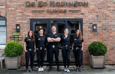
Dr Ed Robinson Clinics
Doctor-led medical aesthetics, skin & laser clinic in Hale, Cheshire
To find a Hyperpigmentation clinic near you, please click here.
Grow Your Practice with Hyperpigmentation
Whether you are already using Hyperpigmentation or considering adding it to your treatment menu, here is how to make the most of it.

Are you a practitioner? If you'd like to link your practice to Hyperpigmentation and become part of ConsultingRoom.com, you can register as a verified specialist using the information on our specialist registration page.
Once verified, your profile can feature Hyperpigmentation alongside the other treatments, conditions and products you support, helping patients searching ConsultingRoom.com find you directly.
Our Preferred Partner:
As more patients research hyperpigmentation online before booking a consultation, having your clinic listed against this condition on ConsultingRoom.com is becoming an increasingly valuable way to reach them. From melasma and post-inflammatory pigmentation to sun damage and age spots, patients turn to pages like this to understand their options before choosing a practitioner.
If your clinic treats hyperpigmentation, our dedicated practitioner resource explains everything you need to know, including how to get listed, showcase your expertise and connect with patients actively searching for treatment.
Find out how being listed here could help grow your clinic and connect you with patients seeking trusted, qualified care.